Rib Cage Muscles Anatomy - 8. Muscles of the Spine and Rib Cage | Musculoskeletal Key - But the cartilages of these ribs are not.
byAdmin-
0
Rib Cage Muscles Anatomy - 8. Muscles of the Spine and Rib Cage | Musculoskeletal Key - But the cartilages of these ribs are not.. The muscular system consists of the skeletal muscles and their associated structures. The rib cage is composed by sternum, costal cartilages, and ribs connected to the thoracic vertebrae. So what parts of the rib cage show up on the surface? Another shoulder positioning muscle that can be observed on. Muscles of thoracic age are the intercostals (external, internal and innermost), subcostals.
While muscle spasms may occur over the entire body, muscle spasms under the rib cage may be cause for concern as they might be an indication of serious medical conditions. But the cartilages of these ribs are not. We study anatomy at the practical anatomy class we study the human body. The ribs are a set of twelve paired bones which form the protective 'cage' of the thorax. But for an anatomy study, it's not.
Rib Cage Diagram With Organs - Human Anatomy Body from www.anatomylibrary99.com Rib cage anatomy and breathing. This is a stereogram, to be viewed in crossview technique. So what parts of the rib cage show up on the surface? Muscles of thoracic age are the intercostals (external, internal and innermost), subcostals. The fibers attach to the rib cage and the pubis of the hip bones. The ribs are a set of twelve paired bones which form the protective 'cage' of the thorax. During normal breathing, contraction of the major inspiratory muscle, the diaphragm, produces both rib cage expansion and a downward movement of the diaphragm. Anterior view of the lungs and ribcage in a transparent female torso stock illustration these pictures of this page are about:human anatomy rib cage muscles.
Volume rendering of a contrast enhanced thoracoabdominal ct scan.
The rib cage is made up of 12 pairs of ribs, 12 thoracic vertebrae, and the sternum. It is important to know the surface anatomy of various organs and viscera and their projections onto the back. Volume rendering of a contrast enhanced thoracoabdominal ct scan. I also discussed the anatomy of false ribs, true ribs and floating ribs and the way they articulate with thoracic vertebrae and how they create the thoracic wall. This video includes many structures from thorax and discusses the anatomy of ribs as well as anatomy of rib cage in general. • raise rib cage for inhaling & depresses rib cage for exhaling. Ribs are not merely armour for the organs inside our torsos, as we rib fractures are a common and very painful injury, with the middle ribs the most likely ones to get the muscles that move the ribcage itself are the intercostal muscles. Various skeletal muscles are attached to the rib cage. This is useful in various procedures as well as for the clinical examination of various body systems. Everyone has nice muscles in ct scanning! This cage protects vital organs and is essential for creating negative pressure to inflate lungs. We study anatomy at the practical anatomy class we study the human body. The ribs joint as follows muscles of the thoracic wall contain those that fill and support the intercostal spaces, those that pass between the sternum and the ribs, and those that cross several.
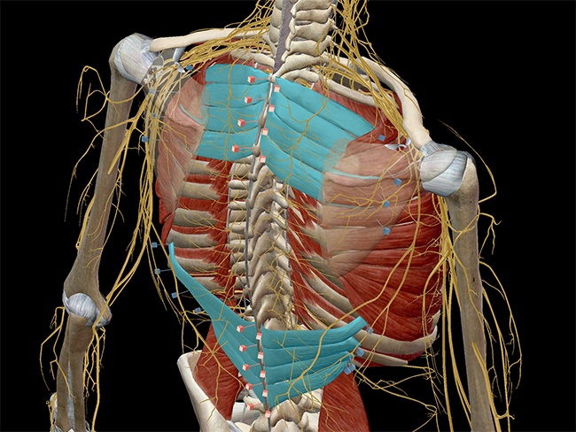
Skeletal muscles attached to the rib cage: The thorax is anatomical structure supported by a skeletal framework (thoracic cage) and contains the the ribs on both the sides complete the cage. Ribs & thoracic cage muscles attachments. Muscle spasms located in the rib cage are often observed in people who strain or overwork their upper body muscles. Serratus posterior superior and inferior.
Learn Muscle Anatomy: Serratus Posterior Superior and Inferior from cdn2.hubspot.net Some extend from above and draw the. Various skeletal muscles are attached to the rib cage. Rib cage anatomy and breathing. On a muscular person when the muscles stretch, we see some of the lower ribs in the front and also in the back. The rib cage is collectively made up of long curved individual bones with joint connections to the spinal vertebrae. Muscles are often named for their primary action. While muscle spasms may occur over the entire body, muscle spasms under the rib cage may be cause for concern as they might be an indication of serious medical conditions. Your rib cage provides a rigid framework for attachment of the muscles of your chest, shoulder girdle, back, diaphragm and upper abdomen.
But the cartilages of these ribs are not.
They are more involved in forced expiration and coughing to forcibly shrink the chest and. The rib cage is collectively made up of long curved individual bones with joint connections to the spinal vertebrae. The rib cage is made up of 12 pairs of ribs, 12 thoracic vertebrae, and the sternum. They are each attached to the ribs. Some extend from above and draw the. For example, flexor, extensor, adductor and abductor are names associated with the action of the muscle. Rib cage anatomy and breathing. Everyone has nice muscles in ct scanning! Muscles are often named for their primary action. Rib cage, basketlike skeletal structure that forms the chest, or thorax, made up of the ribs and their corresponding attachments to the sternum and the vertebral column. The rib cage is composed by sternum, costal cartilages, and ribs connected to the thoracic vertebrae. During normal breathing, contraction of the major inspiratory muscle, the diaphragm, produces both rib cage expansion and a downward movement of the diaphragm. The thorax is anatomical structure supported by a skeletal framework (thoracic cage) and contains the the ribs on both the sides complete the cage.
Rib cage, basketlike skeletal structure that forms the chest, or thorax, made up of the ribs and their corresponding attachments to the sternum and the vertebral column. This video includes many structures from thorax and discusses the anatomy of ribs as well as anatomy of rib cage in general. The ribcage is made to be flexible and springy so the lungs can fill and deflate easily. For a gesture drawing, that's good enough. 1887 human anatomy print of the rib cage and sternum.
Human rib cage, 3/4 front view | Skeleton anatomy, Human ... from i.pinimg.com See more ideas about anatomy, anatomy study, rib cage anatomy. The fibers attach to the rib cage and the pubis of the hip bones. Structure of a typical rib: Another shoulder positioning muscle that can be observed on. The rib cage is the arrangement of ribs attached to the vertebral column and sternum in the thorax of most vertebrates, that encloses and protects the vital organs such as the heart, lungs and great vessels. All three groups of muscles support the rib cage. Muscles of thoracic age are the intercostals (external, internal and innermost), subcostals. Переглядів 46 тис.9 років тому.
The intercostal muscles are the muscles that occupy the 11 intercostal spaces.
The ribcage is made to be flexible and springy so the lungs can fill and deflate easily. They are more involved in forced expiration and coughing to forcibly shrink the chest and. Illustration of thoracic vertebrae showing vertebral body, pedicles, facets, transverse process, rib. Переглядів 46 тис.9 років тому. 1887 human anatomy print of the rib cage and sternum. Muscular system anatomy:muscles of the thoracic cage torso model description. The rib cage is a primarily protective structure, encircling the heart and lungs. Structure and function (6th ed.). Everyone has nice muscles in ct scanning! So what parts of the rib cage show up on the surface? Illustration of rib cage, demonstrating ribs and connection through cartilage to sternum. • raise rib cage for inhaling & depresses rib cage for exhaling. The rib cage, shaped in a mild cone shape and more flexible than most bone sets, is made up of varying elements such as the thoracic vertebra, 12 equally paired ribs, costal cartilage, and held together anteriorly by the sternum.
During normal breathing, contraction of the major inspiratory muscle, the diaphragm, produces both rib cage expansion and a downward movement of the diaphragm rib cage muscles. The ribs joint as follows muscles of the thoracic wall contain those that fill and support the intercostal spaces, those that pass between the sternum and the ribs, and those that cross several.